Growth Prediction Methods
Discuss various methods of studying growth. What are the diagnostic methods you will use to predict class 3 growing malocclusion?
Answer. The following are the two major approaches of studying growth.
- Measurement approach
- Experimental approach.
Measurement Approach
This approach includes techniques that measure certain criteria on living animals/skeletal remains. These techniques are not invasive. Most growth studies on humans are conducted by measurement techniques.
Various measurement techniques can be used on living individuals or the skeletal remains including:
- Craniometry
- Anthropometry
- Cephalometric radiography
- Arcial growth
- Logarithmic spiral
- Finite element analysis.
Craniometry
This is the study of shape and form of human head and the skull. This was a measurement approach to study the growth and was one of the earliest approaches in anthropology. This was used to study the skulls which are found in human skeletal remains. So practice of craniometry consists of precise measurements by using landmarks on the skull. Skull is not the single bone and is made by various interlocked plates. Areas where such bones meet are easily identified and such places form major landmarks on the skull. Distances between various points can be measured and form the base of craniometry. So, in this manner structural model of skull which consists of angles and length between landmarks can be formed and so it is possible to compare one skull with another. Main advantage of craniometry is ability for the precise measurements which can be done on dry skulls. Only cross sections studies can be applied by craniometry.
Anthropometry
It refers to the measurement of a human individual. This is the early tool of physical anthropology and is used to identify and understand the human physical variation and also to correlate with physical and racial psychological traits. Anthropometry consists of systemic measurement of physical properties of human body mainly dimensional descriptors of both the size and shape of the body. Anthropometry uses various landmarks which are used in the study of dry skulls and are measured in the living individuals simply by using the sof tissue points overlying such bony landmarks. Anthropometry can follow the growth of an individual directly and make same measurements repeatedly at different times. This study produces longitudinal data.
Cephalometric Radiography
This technique has contributed majorly in our study of growth and development before it became a routine practice to use the cephalogram for orthodontic diagnosis and planning. Standard cephalometric points are noted on serial radiographs of individuals and compared to analyze the growth changes occurring.
Experimental Approach
This approach includes techniques that may be manipulative and invasive in nature and thus may harm the animal. Such studies are carried out on experimental animals. Experimental methods of study growth include the following:
- Vital staining
- Radioisotopes
- Autoradiography
- Implant radiography.
Vital Staining
- Certain vital stains can be used to determine the sequence and amount of new bone formation as well as specific locations of bone growth by utilizing histologic sections.
- The method involves injecting the dyes that stain the mineralizing tissues.
- These stains get incorporated into the bones and teeth and thus allow the study of changes in bones and teeth.
- Experimental animals are then sacrificed and the mineralizing tissues are studied histologically.
- By this method, detailed analysis of site, amount and rate of growth can be elicited.
- However, this does not allow longitudinal study. Repeated data of the same individual over time cannot be obtained.
- Examples of stains are Alizarin S, procion, tetracycline, trypan blue and florochrome.
Radioisotopes
Radioactive elements can be injected into tissues of experimental animals which get incorporated into the developing bone. Bone growth can be studied tracking the radioactivity emitted by those radioisotopes. For example, calcium 45, technetium 33 (Ca 45, Tc 33).
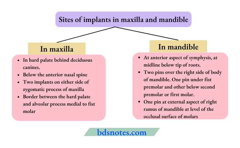
Implant Radiography
- Bjork in 1969 introduced the use of implants to study the bone growth.
- It is an experimental method to study the physical bone growth.
Procedure
- This mainly involves the implanting of small bit of biologically inert alloys inside the growing bone.
- These act as radiographic reference points for the serial radiographic analysis.
- Metallic implants used to study growth are very small mainly 1.5mm in length and 0.5mm in diameter and they are made of tantalum metal.
- These are embedded in various areas of both the maxilla and mandible to study growth of the skull.
Following are the Diagnostic Methods to Predict Class 3 Growing Malocclusion:
Model analysis: In model analysis arch length discrepancy is seen.
Functional analysis: In functional analysis, aberrations are reported in the normal functions such as respiration, swallowing, etc.
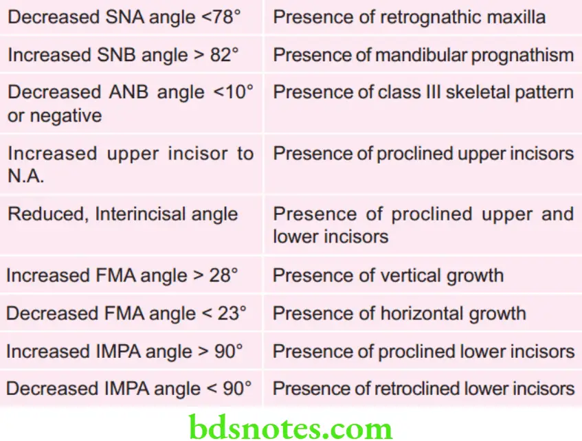
Cephalometrical analysis: In cephalometrical analysis following features are seen.
Leave a Reply