“What is a frenectomy and why is it performed?”
Frenectomy
Frenum
A frenum is a fold of mucous membrane usually with enclosed muscle fibers, that attaches the lips and cheeks to the alveolar mucosa and/or gingiva and underlying periosteum.
“Role of labial frenectomy in correcting diastema”
- A frenum that encroaches the margin of the gingiva may interfere with plaque removal and tensions on this frenum may tend to open the sulcus.
- In these cases, surgical removal of frenum (frenectomy) is indicated.
- High frenum attachment on the lingual surface are uncommon. To correct these without involving the structures in the floor of the mouth, approximately 2 mm of the attachment is separated from the mucosa with a periodontal knife at weekly intervals until the desired level is reached.
- The area is covered with a periodontal pack in the intervals between treatment.
- Frenectomy: It is the complete removal of frenum including its attachment to the bone. It is indicated for correction of abnormal diastema.
- Frenotomy: It is the incision and relation of the frenum to create a zone of attached gingiva between the gingival margin and the frenum.
“Understanding the role of frenectomy in improving oral health”
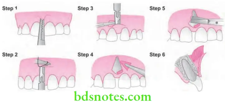
Technique for the Removal of the Frenum (Frenectomy)
- Step 1: After anesthetizing the area, engage the frenum with a hemostat.
- Step 2: Incise along the upper surface of the hemostat, simultaneously make a similar incision along the under surface of the hemostat.
- Step 3: Remove the triangular resected portion of the frenum along with hemostat. This exposes the fibrous connective tissue attachment to bone.
- Step 4: Make a horizontal incision to dissect and separate the fibers attached to bone.
- Step 5: If needed extend the incisions laterally and suture labial mucosa to apical periosteum.
- Step 6: Clean the surgical field and pack with gauze sponges till bleeding stops.
- Step 7: Cover the area by dry aluminum foil and apply periodontal pack
- Step 8: Remove the pack after 2 weeks and repack if needed.
“Importance of studying frenectomy for dental professionals”
“Common challenges in performing frenectomy procedures”
Frenum Attachment And Its Management
Tension Test
Tension test is done to identify whether the width of attached gingiva is adequate or inadequate.
“Steps to differentiate between labial and lingual frenectomy”
Frenum Procedure
This is done by stretching the lip or cheek to demarcate the mucogingival line and to see for any movement of free gingival margin. If the free gingival margin moves during stretching of lips then the attached gingiva is considered to be inadequate and the tension test said to be positive and if gingival margin does not move, test is said to be negative.
“Case studies on outcomes of frenectomy surgeries”
Frenum Significance
- To identify mucogingival line.
- To detect any abnormal frenum.
- To know whether attached gingiva is adequate or inadequate.
Leave a Reply