Advanced Diagnostic Aids In Periodontics
Describe in detail various advanced diagnostic aids.
Or
Write in detail about advanced diagnostic aids in periodontium
Answer. Advanced diagnostic aids are used in periodontics to overcome the problems of conventional diagnostic aids.
Following are the various advanced diagnostic techniques used in periodontology
Advances in Clinical Diagnostic Aids
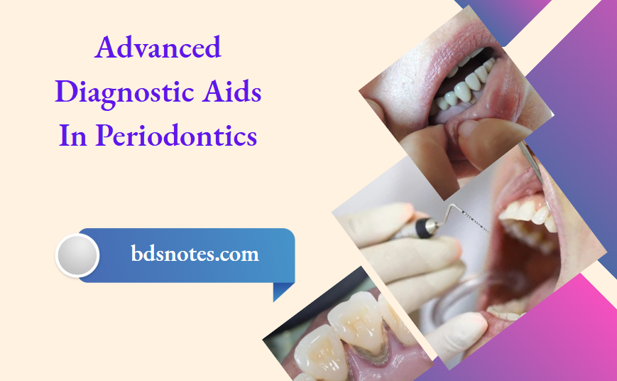
- Periodontal Probes
- Florida probe system: This automated probe system consists of a probe hand piece, digital readout, foot switch, computer interface, and computer. The end of the probe tip is 0.4 mm in diameter. This probe tip reciprocates through a sleeve, and the edge of the sleeve provides a reference by which measurements are made. These measurements are made electronically and transferred automatically to the computer when the foot switch is pressed. Constant probing force is provided by coil springs inside the probe hand piece and digital readout. This probing method combines the advantages of constant probing force with precise electronic measurement and computer storage of data, thus eliminating the potential errors associated with visual reading and the need for an assistant to record the measurements.
- Periotemp: It enables the calculation of temperature differential between the probed pocket and the subgingival temperature. This temperature differential is useful, because it allows consideration of differences in core temperature between individuals. Elevated subgingival site temperature was particularly related to attachment loss in shallow pockets.
- Periodontal Screening and Recording
- It is designed for easier and faster screening and recording of the periodontal status of a patient or a group of population.
- It uses a especially designed probe that has a 0.5 mm ball tip and is color-coded from 3.5 to 5.5 mm.
- Patient’s mouth is divided into six sextants, i.e. maxillary right quadrant, left quadrant and anteriors, mandibular left and right quadrants and anteriors, and at least six points around each tooth is examined.
- The deepest finding is recorded in each sextant according to the following code.
Code 0: In the deepest sulcus of the sextant, the probes colored band remains completely visible, gingival tissue is healthy and does not bleed on gentle probing. No calculus or defective margins are found. These patients require only appropriate preventive care.
Code 1: The colored band remains completely visible in the deepest sulcus of the sextant. No calculus or defective margins are found but some bleeding after gentle probing is found. Treatment for these patients include subgingival plaque removal and appropriate oral hygiene instructions.
Code 2: The probe’s colored band is still completely visible, but there is bleeding on probing. Supragingival or subgingival calculus and/or defective margins are found. Treatment includes plaque and calculus removal, correction of plaque retentive margins of restorations and oral hygiene instructions.
Code 3: The colored band is partially-submerged. This indicates the need for a comprehensive periodontal examination and charting of the affected sextant to determine the necessary treatment plan. lf two or more sextants score code 3, a comprehensive full mouth examination and charting is indicated.
Code 4: The colored band completely disappears in the pocket, indicating a depth greater than 5.5 millimeters. In this case, a comprehensive full mouth periodontal examination, charting and treatment planning are needed.
Code*: When any of the abnormalities are seen, an asterisk (*) is entered, in addition to the code number.
Advances in Radiographic Diagnostic Aids
- Digital Radiography (RVG):
- Digital radiography allows the use of computerized images, which can be stored, manipulated, and corrected for underexposures and overexposures.
- Digital radiography may yield image properties almost equal to conventional radiographs, but through digital storage and processing, diagnostic information can be enhanced.
- Moreover, there is a one-third to half reduction in radiation dose obtained with digital radiographs compared with conventional radiographs.
- Digital intraoral radiography is in a state of rapid development. Sensors as well as computer hardware and software, are continually modified and improved.
- Because of the clear advantage of real or almost-real images that can be improved and the important educational component of online images presented to the patient, it is expected that digital radiography will soon replace conventional radiography in modern daily practice.
- Computer-assisted densitometric image analysis (CADIA)
- In CADIA, a video camera measures the light transmitted through a radiograph, and the signals from the camera are converted into gray-scale images. The camera is interfaced with an image processor and a computer that allow the storage and mathematic manipulation of the images.
- CADIA appears to offer an objective method for following alveolar bone density changes quantitatively over time. Also, compared with digital subtraction analysis.
- CADIA has shown a higher sensitivity and a high degree of reproducibility and accuracy. This technique has also been applied to longitudinal clinical studies.
- Computerized tomography (CT) scan
- Unlike conventional radiography, which is a two dimensional representation of a three-dimensional object. Computed tomography gives an exact picture of the bone levels in coronal, axial and sagittal plane by which all the osseous defects can be visualized accurately.
- Subtraction radiography: In this procedure, two radiographs are taken and the changes are noted depending on the grey levels.
- Digital subtraction radiography
- Digitalization is done before subtraction, i.e. serial radiographs are converted to digital images.
- Digital images are superimposed and are used on a video screen. Light areas indicate bone gain and dark areas indicate bone loss.
- Absorptiometry: A non-radiographic method to analyze the periodontal bone mass changes. It is based on the absorption by bone of a low energy gamma beam, originating from a radioactive source of l25-I. This method has shown to measure bone changes with a high degree of accuracy and precision.
- Photo-densitometric analysis technique: A beam of light is passed onto the radiographic film and the image is shown on an aluminum scale and then it transforms the density readings into millimeter of aluminum equivalents. It is mainly developed to evaluate bone resorption especially in furcation areas. This technique mainly enables the clinician to detect the variations in the bone density that cannot be detected by visual inspection.
- Nuclear medicine bone scan
- It involves the detection of changes in bone metabolism, so it can detect the earliest stage of bone loss.
- A bone seeking radiopharmaceutical diphosphonate compound is injected intravenously and after the waiting period the uptake by the bone is measured by the semiconductor probe radiation detector.
- This technique is of importance because it detects the bone changes before structural alterations occur.
Advances in Microbiological Diagnostic Aids
- Bacterial culture
- Generally, plaque samples are cultivated under an aerobic conditions, and the use of selective and nonselective media, with several biochemical and physical tests allowing the identification of different putative pathogens.
- The main advantage of this method is that the clinician can obtain relative and absolute counts of the cultured species.
- Moreover, it is the only in vitro method able to assess for antibiotic susceptibility of the microbes.
- Direct microscopy
- Specimens are viewed directly under the light. They are of two types:
- Light microscopy: Under this, stained or unstained specimens can be read.
- Gram’s staining: Differentiates Gram-positive and Gram-negative organisms. Gram-positive appears violet.
- Gram-negative appears pink under the micro-scope.
- This may be important because it differentiates between health and disease.
- Dark field and phase contrast microscopy: Fresh, unstained samples are examined. It uses a special condenser in which the light rays are either reflected or refracted off bacterial cell surface. So the outline of the bacterium is dark against the light background in phase contrast microscopy and light against a dark background in dark-field microscopy.
- Advantages of direct microscopy: It is quick, easy and inexpensive means of screening a microbial sample for major morphotypes.
- Disadvantages:
- Inability to identify species.
- Specimens have to be examined as soon as they are collected from the patients.
- Light microscopy: Under this, stained or unstained specimens can be read.
- Specimens are viewed directly under the light. They are of two types:
- Enzymatic methods
- Tannerella forsythia (Tf), Porphyromonas gingivalis (Pg), the small spirochete Treponema denticola (Td), and Capnocytophega species share a common enzymatic profile: all have a trypsin-like enzyme.
- The activity of this enzyme can be measured with the hydrolysis of the colorless substrate N-benzoyl-DLarginine-naphthylamide (BANA).
- When the hydrolysis takes place, it releases the chromophore 5-naphthylamide, which turns orange red when a drop of fast garnet is added to the solution. Diagnostic kits have been developed using this reaction for the identification of this bacteria profile in plaque isolates (Perioscan).
- Immunodiagnostic methods
- ELISA
- In this, bacterial antigens are incubated in a well, on a plastic plate to allow coating by the material. After washing to remove the free antigen, the plates are ready for tests. Samples containing suspected antibodies and controls are then incubated in separate wells to allow antibodies bind the antigen on the surface of the wells. After washing to remove unbound serum components, antisera to the antibody is conjugated to either alkaline phosphatase or horseradish peroxidase then incubated in the wells. A positive reaction is visualized by addition of a chromogen which changes from a colorless to colored solution.
- Latex agglutination:
- It is based on the binding of protein to latex. Latex beads are coated with species specific antibody and when these beads come in contact with the microbial cell surface. Antigens crosslinking occurs and its clumping/agglutination is made visible within 2 to 5 minutes.
- Direct and Indirect immunofluorescent assay:
- This method permits the identification of specific bacteria in bacterial smears.
- ELISA
- Direct immunofluorescence:
- Antiserum to a microorganism is conjugated to fluorescein.
- The conjugate is incubated on a clinical smear containing the microorganisms and then washed off.
- The antigen-antibody reactions take place and organism is visualized by its fluorescent outline, when observed under a fluorescent microscope, if the microorganism is not present, it appears dark with no fluorescence.
- Indirect immunofluorescence: It is a two step procedure. Antiserum to the microorganism is incubated on the clinical smear and washed off, then a conjugate of a fluorescent dye and an antiserum to the first antisera are incubated and then washed off.
- Flow cytometry: This is for rapid identification of oral bacteria. This involves labeling bacterial cells from a patient plaque sample with both species specific antibody and a second fluorescein conjugated antibody. The suspension is then introduced into the flow cytometer, which separates the bacterial cells into an almost single cell suspension by means of a laminar flow through a narrow tube. After incubation, the cells are passed through a focused laser beam. The cells then scatter the light at low and wide angles, and the fluorescent emission can be measured by appropriate detectors.
Advances in Characterizing Host Response Aids
- GCF assessment (Periotron)
- At present, the most common way to assess GCF is using the Periotron. This electronic device measures the change in capacitance across the wetted strip, and this change is converted to a digital readout, which can be correlated to the volume of GCF. Researchers have established that the Periotron 6000 achieves the easiest and quickest measurement and shows high correlation with other clinical gingival indices.
- Evaluation of inflammatory mediators and products
- Cytokines are potent local mediators of inflammation that are produced by a variety of cells. Cytokines that are present in GCF and have been investigated as potential diagnostic markers include tumor necrosis factor alpha (TNF-a), interleukin-1a (IL-1a, interleukin-1b, Interleukin-6 (IL-6), and interleukin-8 (IL-8). lL-6, and TNF-a are cytokines produced by a variety of cells at inflamed sites. They are potent immunoregulatory molecules with a variety of biologic effects, including metalloproteinase stimulation and bone resorption; therefore they seem good candidates for markers of disease progression.
- Host-derived enzymes: Enzymes such as matrix metalloproteinases, alkaline phosphatases, tissue inhibitors of matrix metalloproteinases, aspartase aminotransferase are used as potential diagnostic markers.
- Tissue breakdown products: One of the major features of periodontitis is the destruction of collagen and extracellular matrices. The connective tissues of the periodontium are composed of fibrous elements, including proteins such as collagen and elastin, and nonfibrous components, including a variety of glycoproteins (laminin, fibronectin, proteoglycans) as well as minerals, lipids, water, and tissue-bound growth factors. The extracellular matrix of the periodontium is composed of a diverse number of macromolecules; the predominant one is collagen, and the other components include proteoglycan (versican, decorin, biglycan, syndecan) and noncollagen proteins (elastin, fibronectin, laminin, osteocalcin, osteopontin, bone sialoprotein, osteonectin, tenascin). All these matrix components are theoretically detectable and potentially informative in terms of their clinical diagnostic utility.
Leave a Reply