Sublingual Dermoid Cyst In Young Child
Sublingual Dermoid Cyst
Answer. Sublingual Dermoid Cyst is a type of congenital sequestration dermoid cyst.
The cyst is formed by the inclusion of the surface ectoderm at the fusion line of two mandibular arches.
Sublingual Dermoid Cyst Pathology
- The cyst is lined by squamous epithelium.
- The wall of the cyst contains hair follicles, sweat, and sebaceous glands.
- Cyst contains cheesy material.
- It never contains hair.
Sublingual Dermoid Cyst In Children
Read and learn More Cysts: Types, Causes, Symptoms, and Treatment
“Can Sublingual Dermoid Cyst Cause Breathing Problems”
Sublingual Dermoid Cyst Types may be:
- Median variety: It is derived from epithelial cell rests at the level of fusion of two mandibular arches.
It may be supramylohyoid or infra mylohyoid.
It is located between two genial muscles, about the mylohyoid muscle.
It is a midline swelling that is smooth, soft, cystic, nontransilluminant. - Lateral variety: It develops about the submandibular duct, lingual nerve, and stylohyoid ligament.
It is derived from the first branchial arch.
It forms a swelling in the lateral aspect of the floor of the mouth.

Pediatric Sublingual Dermoid Cyst
“Effective Ways To Manage Sublingual Dermoid Cyst Recovery”
It also may be:
- Supra mylohyoid type.
- Infra mylohyoid type.
Sublingual Dermoid Cyst Clinical Features
- It occurs in young children between the ages of 10 to 12 years.
- Congenital, painless, and digitally palpable swelling in the floor of the mouth.
- Swelling is soft and cystic
- The fluctuation test is positive.
- The transillumination test is negative as it contains thick, cheesy, sebaceous material.
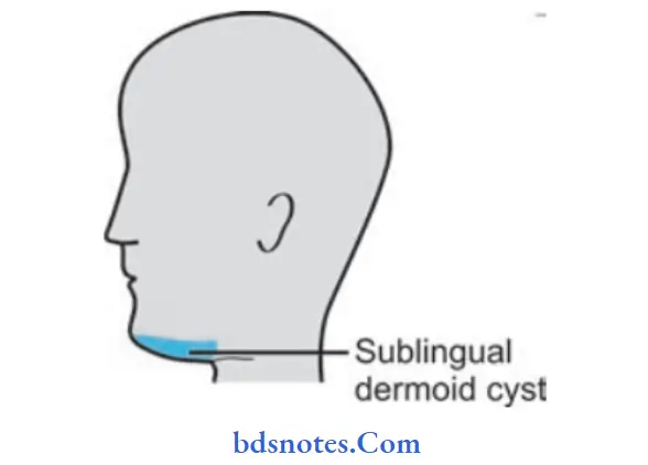
- Swelling may often attain a large size presenting both sublingually, intraorally, and midline submentally on the external side.
- Occasionally it can lead to trismus, dysphagia, pain, and odynophagia.
Congenital Sublingual Dermoid Cyst
“What Tests Diagnose Sublingual Dermoid Cyst In Kids”
Sublingual Dermoid Cyst Differential Diagnosis
- Ranula: When the sublingual dermoid cyst is in the midline at the floor of the mouth and above the mylohyoid muscle ranula is considered a differential diagnosis.
Ranula is blue in color and brilliantly translucent. - Thyroglossal cyst: It is to be taken in the differential diagnosis when the sublingual dermoid cyst is below the mylohyoid muscle.
The thyroglossal cyst moves up with deglutition whereas the sublingual dermoid cyst does not.
Sublingual Dermoid Cyst In Infants
Sublingual Dermoid Cyst Treatment
Excision is done through an intra-oral approach usually; large cystic tending under the geniohyoid muscle may require external approach.
Leave a Reply