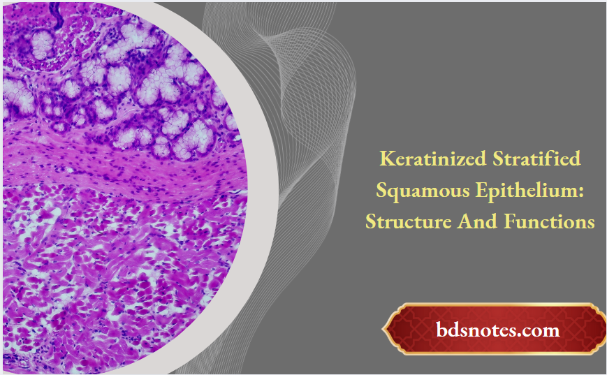
Keratinized Stratified Squamous Epithelium
Write a short note on the stratified squamous keratinized epithelium.
Answer.
Stratified Squamous Keratinized Epithelium Characteristic features
They are same as that of stratified squamous nonkeratinized epithelium except that the surface cells undergo keratinization (i.e. cells become dead, lose their nucleus and get filled with keratin).
Keratinized Stratified Squamous Epithelium Definition

“Functions Of Keratinized Stratified Squamous Epithelium”
Stratified Squamous Keratinized Epithelium Function
Protection (i.e. prevents wear and tear)
Keratinized Epithelium Structure
Stratified Squamous Keratinized Epithelium Sites
- Skin
- Vestibule of nose
- External auditory meatus
- Lower part of anal canal
Keratinized stratified squamous epithelium is an important type of tissue that covers and protects various surfaces of the body. It’s made up of multiple layers of cells, with the topmost layer being tough and filled with keratin. This structure plays a key role in safeguarding our skin and other areas from damage, dehydration, and infection. Understanding its layers and functions can help us appreciate how our body stays protected against the environment.
“Where Is Keratinized Stratified Squamous Epithelium Found”
- Keratinized stratified squamous epithelium consists of several layers of cells, with the outermost layer being keratinized for protection.
- Keratinized stratified squamous epithelium is primarily found in the epidermis of the skin, as well as in areas like the oral cavity and parts of the tongue.
- The layers include the stratum basale, spinosum, granulosum, lucidum, and corneum, each with specific roles.
- This tissue acts as a barrier to prevent water loss and protects against environmental threats.
- Clinical issues can arise from changes in this epithelium, such as skin disorders and the process of metaplasia.
Defining Keratinized Stratified Squamous Epithelium
Okay, so let’s talk about keratinized stratified squamous epithelium. It’s a mouthful, I know, but breaking it down makes it easier. Think of it as a type of tissue that’s built for protection. It’s found in places that take a beating, like your skin. It’s made up of layers of cells, and the outermost layer is toughened up with keratin, a protein that makes it waterproof and resistant to damage. It’s like the body’s natural armor.
Function Of Keratinized Stratified Squamous Epithelium
Characteristics of Keratinized Cells
Keratinized cells are pretty special. They start out as normal cells in the deeper layers, but as they move towards the surface, they fill up with keratin. This process changes their structure and function. They become flat, tough, and eventually die, forming a protective layer. It’s kind of like they sacrifice themselves for the good of the body. Here’s a few things that happen:
- Cells produce lots of keratin.
- The nucleus and other organelles break down.
- Cells become tightly packed and flattened.
Importance of Keratinization
Why is keratinization so important? Well, it’s all about protection. The keratin layer acts as a barrier against all sorts of things, like bacteria, viruses, and physical damage. It also helps to prevent water loss, which is crucial for keeping our bodies hydrated. Without keratinization, we’d be much more vulnerable to the environment. Think of it like this:
Keratinization is like the body’s sealant, keeping the good stuff in and the bad stuff out. It’s a simple process with a huge impact on our health and well-being.
“How Does Keratinized Stratified Squamous Epithelium Protect The Body”
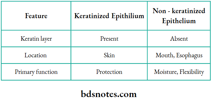
Comparison with Non-Keratinized Epithelium
So, what’s the difference between keratinized and non-keratinized epithelium? The main difference is the presence of that tough keratin layer. Non-keratinized epithelium is found in places that need to stay moist, like the lining of your mouth or esophagus. It doesn’t have the same level of protection as keratinized epithelium, but it’s more flexible and can secrete mucus. Here’s a quick comparison:
Location Of Keratinized Stratified Squamous Epithelium
Basically, it all comes down to location and function. The body uses different types of epithelium in different places, depending on what kind of job needs to be done. For example, the epidermis of skin needs to be tough, while the lining of the mouth needs to be moist.
Layers of Keratinized Stratified Squamous Epithelium
Alright, let’s break down the layers of this tough tissue. Think of it like a fortress, each layer with its own job to do. It’s not just a random pile of cells; there’s a specific order and purpose to each level.
Stratum Basale
This is the foundation, the bottom layer. It’s where new cells are born. These cells are constantly dividing, pushing older ones up to the surface. You can think of it as the engine room of the whole operation. It’s attached to the basement membrane, providing structural support and nutrients. It’s also called the stratum germinativum.
“Is Keratinized Stratified Squamous Epithelium Waterproof”
Stratum Spinosum
Above the basale, we’ve got the spinosum. These cells are a bit bigger and have spiny projections that connect them to each other. These spines are actually desmosomes, which are like little rivets holding the cells together. They provide strength and flexibility to the tissue. The cells here have migrated from the stratum basale.
Stratum Granulosum
Next up is the granulosum. This is where things start to get serious. The cells here are filled with granules that contain lipids and proteins. These granules help to form a waterproof barrier, preventing water loss from the body. The cells are starting to flatten out and die as they move towards the surface. It’s a critical step in the process of keratinization.
Stratum Lucidum
This layer is only found in thick skin, like on the palms of your hands and soles of your feet. It’s a clear, translucent layer of dead cells. These cells are packed with eleidin, a precursor to keratin. It provides extra protection and durability in areas that experience a lot of wear and tear. It’s not present in all types of keratinized stratified squamous epithelium.
Keratinized vs Non-Keratinized Epithelium
Stratum Corneum
Finally, we reach the top – the stratum corneum. This is the outermost layer, and it’s made up of dead, flattened cells filled with keratin. These cells are constantly being shed and replaced by new cells from below. It’s the main barrier against the outside world, protecting us from damage, infection, and dehydration. It’s like a shield, taking all the hits so the layers underneath stay safe.
So, there you have it – the five layers of keratinized stratified squamous epithelium. Each layer plays a vital role in protecting our bodies. From the actively dividing cells at the bottom to the tough, dead cells at the top, it’s a complex and fascinating system.
Here’s a quick rundown:
- Stratum Basale: Cell division
- Stratum Spinosum: Strength and flexibility
- Stratum Granulosum: Waterproof barrier
Functions of Keratinized Stratified Squamous Epithelium
Protection Against Environmental Damage
Keratinized stratified squamous epithelium is like your body’s first line of defense. Think of it as a tough shield. Its primary job is to protect against all sorts of environmental hazards. This includes things like UV radiation from the sun, physical abrasion, and even chemical exposure. The tightly packed layers of cells, especially the stratum corneum, create a barrier that’s hard to penetrate. It’s not perfect, but it does a pretty good job of keeping the bad stuff out.
“Role Of Keratin In Keratinized Stratified Squamous Epithelium”
Barrier to Water Loss
One of the most important functions of this epithelium is preventing dehydration. The keratin in the cells is hydrophobic, meaning it repels water. This helps to minimize water loss from the underlying tissues. Without this barrier, we’d dry out super fast! It’s like having a built-in moisturizer, but way more effective. This is especially important in areas like the skin, where exposure to the air can quickly lead to water evaporation. Think of it like this:
- Keratin acts as a water repellent.
- Multiple layers provide a thicker barrier.
- Cell junctions further seal the tissue.
The barrier function is so effective that it also prevents excessive water absorption from the environment. This is why you don’t swell up like a sponge when you take a bath.
Role in Sensation
While the keratinized layer is dead, it’s closely associated with sensory nerve endings in the underlying layers. These nerve endings allow us to feel things like pressure, pain, and temperature. The epithelium itself doesn’t “feel”, but it transmits stimuli to the nerves below. It’s like a messenger service, relaying information from the outside world to our nervous system. The density of these nerve endings varies depending on the location, which is why some areas of the skin are more sensitive than others. For example, fingertips have a much higher density of nerve endings than the skin on your back. This allows for fine motor skills and detailed tactile perception. The epithelial tissues are essential for this process.
Locations of Keratinized Stratified Squamous Epithelium
Keratinized stratified squamous epithelium is strategically located in the body to provide protection against abrasion, water loss, and other environmental stressors. It’s found in areas that endure a lot of friction or need a strong barrier.
Epidermis of the Skin
The epidermis, the outermost layer of our skin, is the most prominent location of this type of epithelium. It’s designed to withstand constant wear and tear, protecting underlying tissues from injury, infection, and dehydration. Think about how much your hands and feet are used every day – that’s keratinized stratified squamous epithelium at work!
Oral Mucosa
Not all of the inside of your mouth is the same. Areas of the oral mucosa that experience a lot of friction, like when you’re chewing, are lined with keratinized stratified squamous epithelium. This helps them hold up better.
Keratinized Epithelium Characteristics
Gingiva and Hard Palate
Specifically, the gingiva (gums) and the hard palate (the roof of your mouth) are prime examples. These areas are subject to considerable mechanical stress during chewing. The keratinized layer provides a tough, protective surface. It’s interesting to note that the presence and degree of keratinization can vary in different parts of the oral cavity, depending on the functional demands of each area.
The distribution of keratinized stratified squamous epithelium is a great example of how structure follows function in the human body. The body places this tough tissue exactly where it’s needed most to withstand physical stress and maintain a barrier against the outside world.
“Understanding Keratinized Stratified Squamous Epithelium In Dry Skin”
Clinical Significance of Keratinized Stratified Squamous Epithelium
Implications in Skin Disorders
Keratinized stratified squamous epithelium is super important for skin health, so when things go wrong with it, you see a bunch of different skin problems. For example, in psoriasis, there’s too much keratinization, which leads to scaly, itchy skin. Eczema, on the other hand, can involve a breakdown of this epithelial barrier, making the skin more vulnerable to irritants and allergens. Understanding how this epithelium functions normally is key to figuring out what’s happening in these skin disorders and how to treat them.
Role in Wound Healing
This type of epithelium plays a big role in how wounds heal. When you get a cut or scrape, the keratinized stratified squamous epithelium works to close the gap. Here’s a quick rundown:
- First, cells from the edges of the wound start to migrate and proliferate.
- Then, they lay down new tissue to fill in the damaged area.
- Finally, the new epithelium matures and keratinizes, restoring the skin’s protective barrier.
If this process is disrupted, it can lead to chronic wounds or excessive scarring. So, keeping the wound clean and protected is important for proper epithelial regeneration.
Metaplasia and Its Effects
Sometimes, under chronic irritation or stress, keratinized stratified squamous epithelium can change into a different type of epithelium through a process called metaplasia. This is like the body trying to adapt, but it can have some not-so-great consequences. For instance, in the esophagus, long-term acid reflux can cause the normal squamous epithelium to be replaced by columnar epithelium, which is what you see in Barrett’s esophagus. This change increases the risk of esophageal cancer.
Comparative Anatomy Of Epithelial Types
Differences with Simple Squamous Epithelium
Okay, so let’s talk about how keratinized stratified squamous epithelium stacks up against simple squamous epithelium. Simple squamous is like the minimalist of the epithelium world – a single layer of flat cells. Think of it as a delicate covering, perfect for places where stuff needs to pass through easily, like in the lungs or blood vessels. On the other hand, keratinized stratified squamous is the body’s heavy-duty armor. It’s got multiple layers, and the top layer is packed with keratin, making it tough and waterproof. It’s all about protection, not permeability. You’ll find simple squamous epithelium where quick diffusion is key, while the stratified version is where you need a barrier.
“Treating Damage To Keratinized Stratified Squamous Epithelium In The Skin”
Contrasts with Stratified Cuboidal Epithelium
Now, let’s throw stratified cuboidal epithelium into the mix. This type is made up of multiple layers of cube-shaped cells. It’s not as common as the other two, but you’ll find it in places like sweat glands and salivary glands. It’s more about secretion and protection than absorption or diffusion. The big difference is the cell shape and the level of protection. Keratinized stratified squamous is way tougher because of the keratin, and it’s designed to withstand a lot of wear and tear. Stratified cuboidal is more about lining ducts and providing a bit of a barrier, but it’s not nearly as robust.
Histology Of Keratinized Epithelium
Unique Features of Pseudostratified Epithelium
Finally, there’s pseudostratified epithelium. This one’s a bit of a trickster because it looks like it has multiple layers, but it’s really just one. The cells are different sizes, so their nuclei end up at different levels, making it look stratified. You often find it with cilia, like in the respiratory tract, where it helps move mucus. Here’s a quick rundown:
- Keratinized stratified squamous: Multiple layers, flat cells, keratinized, protection.
- Simple squamous: Single layer, flat cells, diffusion.
- Stratified cuboidal: Multiple layers, cube-shaped cells, secretion and protection.
- Pseudostratified: Single layer (looks like multiple), different cell heights, often ciliated, mucus movement.
So, while keratinized stratified squamous epithelium is all about tough protection, the other types are specialized for different jobs, like diffusion, secretion, or moving stuff around. It’s all about form following function in the amazing world of tissues.
“Preventing Complications From Damaged Keratinized Stratified Squamous Epithelium”
Histological Examination of Keratinized Stratified Squamous Epithelium
Microscopic Structure
When you look at keratinized stratified squamous epithelium under a microscope, the most striking thing is the layered arrangement. You’ll see several distinct layers, each with cells in different stages of development. The deepest layer, the stratum basale, is where new cells are born. As they move towards the surface, they change shape and accumulate keratin. The outermost layer, the stratum corneum, is made up of dead, flattened cells packed with keratin, providing a tough, protective barrier. The structural diversity is pretty amazing.
Staining Techniques
To really see the details, we use stains. Hematoxylin and eosin (H&E) is a common choice. Hematoxylin stains the nuclei a dark blue or purple, while eosin stains the cytoplasm and extracellular proteins pink. This helps differentiate the different layers and cell types. Other stains, like Masson’s trichrome, can highlight collagen fibers in the underlying connective tissue. Periodic acid–Schiff (PAS) stain can be used to identify glycogen and other carbohydrates in the cells. Here’s a quick rundown:
- H&E: General structure, nuclei (blue), cytoplasm (pink)
- Masson’s Trichrome: Collagen (blue or green), muscle fibers (red)
- PAS: Carbohydrates (magenta)
Identifying Features Under Microscope
So, what are the key things to look for? First, the thickness of the epithelium is a big clue. Keratinized stratified squamous epithelium is generally quite thick, especially in areas like the skin. You’ll also notice the distinct layers: stratum basale, stratum spinosum, stratum granulosum, stratum lucidum (sometimes), and stratum corneum. The stratum corneum is the most obvious, with its flattened, anucleated cells. The presence of keratohyalin granules in the stratum granulosum is another important feature. And of course, the overall arrangement of cells, with squamous cells on the surface and more cuboidal or columnar cells at the base, is characteristic.
It’s important to remember that the appearance can vary depending on the location and the specific staining technique used. But by focusing on the layered structure, the presence of keratin, and the distinct cell types in each layer, you can confidently identify keratinized stratified squamous epithelium under the microscope.
“Dealing With Cracks In Keratinized Stratified Squamous Epithelium”
Wrapping It Up
In summary, keratinized stratified squamous epithelium plays a vital role in protecting our bodies. It’s the tough outer layer of our skin and is also found in areas like the gums and the roof of the mouth. This type of tissue is built with several layers, which helps it withstand wear and tear. The process of keratinization, where cells move up and become filled with keratin, is key to its protective abilities. Understanding this tissue can help us appreciate how our bodies defend against damage and keep moisture in. So next time you think about your skin, remember the hard work that keratinized stratified squamous epithelium does every day!
Histology Of Keratinized Epithelium
Frequently Asked Questions
Question 1. What Is Keratinized Stratified Squamous Epithelium?
Answer: Keratinized stratified squamous epithelium is a type of tissue made up of many layers of flat cells. The top layer is tough and filled with a protein called keratin, which helps protect the skin.
Question 2. Why Is Keratinization Important?
Answer: Keratinization is important because it helps create a barrier that protects our skin from damage, germs, and water loss.
Question 3. How Does Keratinized Epithelium Differ From Non-Keratinized Epithelium?
Answer: The main difference is that keratinized epithelium has a tough outer layer of dead cells filled with keratin, while non-keratinized epithelium does not have this layer and is usually found in moist areas.
“Understanding The Structure Of Keratinized Stratified Squamous Epithelium”
Question 4. Where Can We Find Keratinized Stratified Squamous Epithelium In The Body?
Answer: You can find keratinized stratified squamous epithelium mainly in the outer layer of the skin (epidermis), and in parts of the mouth like the gums and hard palate.
Question 5. What Are The Functions Of This Type Of Epithelium?
Answer: This type of epithelium protects against damage from the environment, helps keep moisture in, and plays a role in sensation.
Question 6. What Happens If There Are Problems With Keratinized Stratified Squamous Epithelium?
Answer: If there are issues with this type of epithelium, it can lead to skin disorders, affect how wounds heal, and sometimes cause changes in the type of cells present, known as metaplasia.
Leave a Reply