• Skip to main content
  • Skip to secondary menu
  • Skip to primary sidebar
  • About Us
  • Terms of Use
  • Privacy Policy
  • Disclaimer
  • Contact Us
  • Sitemap

BDS Notes

BDS notes, Question and Answers

  • Public Health Dentistry
  • Periodontics
  • Pharmacology
  • Pathology
  • Orthodontics
    • Anchorage In Orthodontics
    • Mandibular Growth, Functional Matrix
    • Retention and Relapse
  • General Surgery
    • Cysts: Types, Causes, Symptoms
    • Maxillofacial Fractures, Disorders, and Treatments
    • Lymphatic Disorders
    • Neurological and Facial Disorders
  • Temporal And Infratemporal Regions
    • Spinal and Neuroanatomy
  • Dental Materials
    • Dental Amalgam
Home » Spiral Organ of Corti: Structure, Function, and Its Role in Hearing

Spiral Organ of Corti: Structure, Function, and Its Role in Hearing

February 11, 2025 by Kristensmith Taylor Leave a Comment

Organ Of Corti

Write a short note on the spiral organ of Corti.
Answer.

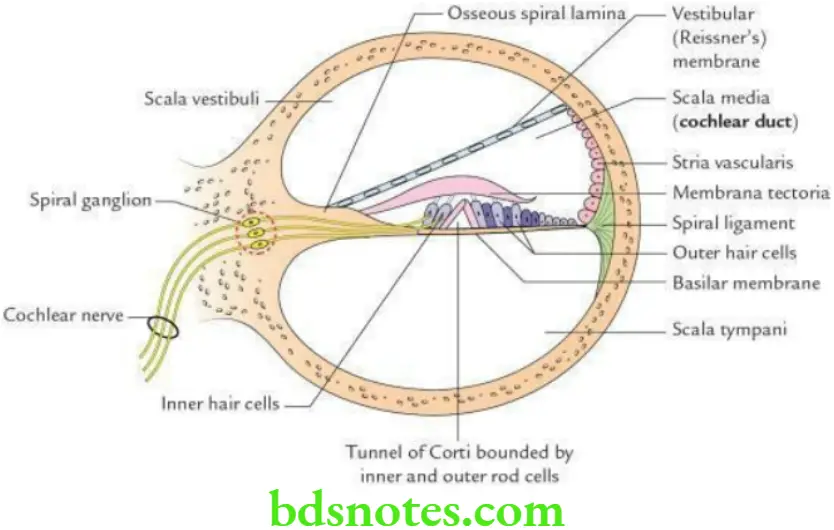
It is an end organ of hearing, located on the basilar membrane of the cochlear duct.

Spiral Organ of Corti Components Microscopically, the organ of Corti consists of five components:

“Importance Of The Organ Of Corti In Sound Perception”

  • Basilar membrane: A fibrous membrane that extends from the osseous spiral lamina to the outer wall of the cochlear duct.
  • Tunnel of Corti: Formed by inner and outer rod cells and contains corticolymph.
  • Hair cells are receptor cells of hearing located on the basilar membrane. These cells bear stereocilia and form the most important component of the spiral organ of Corti. They are divided into inner and outer hair cells.
    The hair cells perform the following functions:

    • Detect movements of endolymph
    • Detect vibrations of the basilar membrane
    • Transfer vibrations into nerve impulses to the cochlear nerve

“Best Ways To Understand The Structure Of The Organ Of Corti”

  • Supporting cells: The inner hair cells are supported by phalangeal cells, while the outer supporting cells are called Henson’s cells.
  • Membrana tectoria: It is a gelatinous membrane that overlies the hair cells. The shearing force between the hair cells and tectorial membrane stimulates the hair cells.

Head And Neck Ear and orbit Spiral organ of corti as seen in a section through the cochlear duct

“Understanding The Role Of Hair Cells In The Organ Of Corti”

Innervation of the organ of Corti The hair cells are innervated by the peripheral processes of bipolar neurons of the spiral ganglion located within the modiolus near the base of the spiral lamina. There are two types of neurons:

Type 1 neurons: They are myelinated and afferent. They innervate inner hair cells and are responsible for auditory sensation.

Type 2 neurons: They are unmyelinated and efferent from the contralateral superior olivary nucleus. They innervate outer hair cells and are responsible for auditory discrimination.

Filed Under: Head And Neck

Reader Interactions

Leave a Reply Cancel reply

Your email address will not be published. Required fields are marked *

Primary Sidebar

Recent Posts

  • Branchial Cleft Cyst: Background, Pathophysiology, Etiology
  • Maxillary Nerve: Origin, Course, And Branches
  • The Father Of Anatomy And A Great Anatomist Herophilus
  • Bone Structure – Anatomy
  • The External Carotid Artery: Anatomy, Branches, And Functions
  • Occipitofrontalis Muscle
  • Superficial Temporal Artery
  • Platysma Muscle
  • Cartilage
  • Cauda Equina And Conus Medullaris Syndromes
  • Subcutaneous Injections And Device Management
  • Types Of Circulation: Pulmonary, Systemic, And Portal
  • Hierarchical Organization Of Skeletal Muscle Tissue
  • Elastic Cartilage Histology Short Note For Medical Exams
  • Cellular Organelles And Structure
  • The Golgi Apparatus – The Cell
  • The Cytoplasmic Inclusions Of Certain Plant Cells
  • Dental Abscess
  • Laser Surgery
  • Our Facial Muscles And Their Functions

Copyright © 2026 · Magazine Pro on Genesis Framework · WordPress · Log in