• Skip to main content
  • Skip to secondary menu
  • Skip to primary sidebar
  • About Us
  • Terms of Use
  • Privacy Policy
  • Disclaimer
  • Contact Us
  • Sitemap

BDS Notes

BDS notes, Question and Answers

  • Public Health Dentistry
  • Periodontics
  • Pharmacology
  • Pathology
  • Orthodontics
    • Anchorage In Orthodontics
    • Mandibular Growth, Functional Matrix
    • Retention and Relapse
  • General Surgery
    • Cysts: Types, Causes, Symptoms
    • Maxillofacial Fractures, Disorders, and Treatments
    • Lymphatic Disorders
    • Neurological and Facial Disorders
  • Temporal And Infratemporal Regions
    • Spinal and Neuroanatomy
  • Dental Materials
    • Dental Amalgam
Home » Mastoid Process: Location, Anatomy And Muscle Attachments

Mastoid Process: Location, Anatomy And Muscle Attachments

February 10, 2025 by Kristensmith Taylor Leave a Comment

Mastoid Process: Location, Anatomy And Muscle Attachments

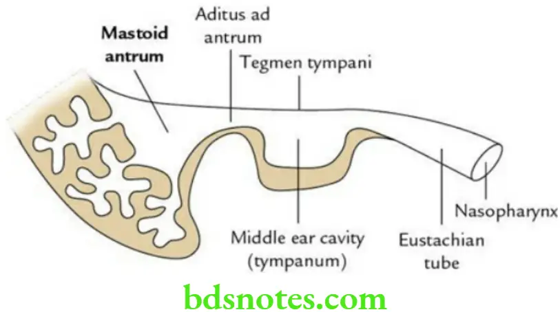
Write a short note on mastoid antrum.
Answer.

  • The mastoid antrum is an air space in the mastoid portion of the temporal bone.
  • Mastoid Antrum communicates anteriorly with the tympanic cavity through aditus ad antrum (entrance to the mastoid antrum).

“Importance Of The Mastoid Process In Skull Anatomy”

Head And Neck Ear and orbit Mastoid antrum as seen in section along the long axis of petromastoid bone

“Role Of The Mastoid Process In Muscle Attachment”

Mastoid Antrum Boundaries

Roof is formed by tegmen tympani.

The lateral wall is formed by a plate of bone about 1.5 cm thick just deep into a supramental triangle.

The posterior wall is formed by a thin plate of bone that separates it from the sigmoid sinus.

“Understanding The Role Of The Mastoid Process In Skull Anatomy”

Mastoid Antrum Functions

  • Provides resonance to the voice.
  • Acts as an acoustic insulator and provides protection to the middle ear from physical damage.
  • Acts as sound receptor.

“Comprehensive Overview Of The Mastoid Process And Its Significance”

Mastoid Antrum Applied anatomy

  • Mastoid air cells are a major contributor to the middle ear inflammatory disease.
  • The lateral wall of the antrum is approached for surgery, through the supramental triangle.

Filed Under: Head And Neck

Reader Interactions

Leave a Reply Cancel reply

Your email address will not be published. Required fields are marked *

Primary Sidebar

Recent Posts

  • Branchial Cleft Cyst: Background, Pathophysiology, Etiology
  • Maxillary Nerve: Origin, Course, And Branches
  • The Father Of Anatomy And A Great Anatomist Herophilus
  • Bone Structure – Anatomy
  • The External Carotid Artery: Anatomy, Branches, And Functions
  • Occipitofrontalis Muscle
  • Superficial Temporal Artery
  • Platysma Muscle
  • Cartilage
  • Cauda Equina And Conus Medullaris Syndromes
  • Subcutaneous Injections And Device Management
  • Types Of Circulation: Pulmonary, Systemic, And Portal
  • Hierarchical Organization Of Skeletal Muscle Tissue
  • Elastic Cartilage Histology Short Note For Medical Exams
  • Cellular Organelles And Structure
  • The Golgi Apparatus – The Cell
  • The Cytoplasmic Inclusions Of Certain Plant Cells
  • Dental Abscess
  • Laser Surgery
  • Our Facial Muscles And Their Functions

Copyright © 2026 · Magazine Pro on Genesis Framework · WordPress · Log in