• Skip to main content
  • Skip to secondary menu
  • Skip to primary sidebar
  • About Us
  • Terms of Use
  • Privacy Policy
  • Disclaimer
  • Contact Us
  • Sitemap

BDS Notes

BDS notes, Question and Answers

  • Public Health Dentistry
  • Periodontics
  • Pharmacology
  • Pathology
  • Orthodontics
    • Anchorage In Orthodontics
    • Mandibular Growth, Functional Matrix
    • Retention and Relapse
  • General Surgery
    • Cysts: Types, Causes, Symptoms
    • Maxillofacial Fractures, Disorders, and Treatments
    • Lymphatic Disorders
    • Neurological and Facial Disorders
  • Temporal And Infratemporal Regions
    • Spinal and Neuroanatomy
  • Dental Materials
    • Dental Amalgam
Home » Understanding The Carotid Sheath: Anatomy, Formation, And Clinical Relevance

Understanding The Carotid Sheath: Anatomy, Formation, And Clinical Relevance

February 11, 2025 by Kristensmith Taylor Leave a Comment

Understanding The Carotid Sheath: Anatomy, Formation, And Clinical Relevance

Write a short note on the carotid sheath.
Answer.

Formation

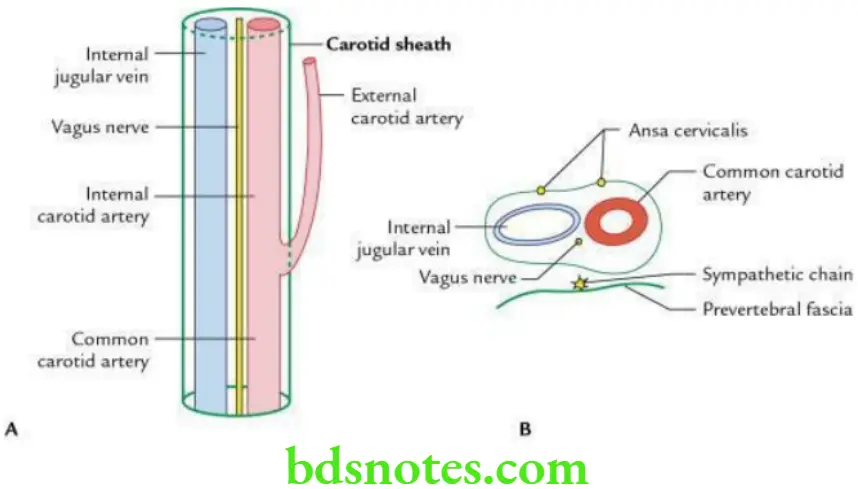
  • It is formed by the condensation of fibroareolar tissue around common and internal carotid arteries and internal jugular vein.
  • It extends from the base of the skull to the arch of the aorta.
  • Its anterior wall is connected to the pretracheal fascia while its posterior wall is connected to the prevertebral fascia.

“Risk Factors For Complications Involving The Carotid Sheath”

Head And Neck Side front and back of the neck Carotid sheath

“Importance Of The Carotid Sheath In Protecting Neck Structures”

Thickness It is thick around common and internal carotid arteries, and thin around the internal jugular vein to allow the free expansion of the vein during increased venous return.

Relations

Anteriorly: Ansa cervicalis is embedded in the wall or sheath.

Posteriorly: The sympathetic trunk is present behind the sheath.

“Understanding The Role Of The Carotid Sheath In Neck Protection”

Contents

  • Common and internal carotid arteries
  • Internal jugular vein
  • Vagus nerve

Applied anatomy It is frequently exposed in block dissection of the neck during surgical removal of the deep cervical lymph nodes.

Filed Under: Head And Neck

Reader Interactions

Leave a Reply Cancel reply

Your email address will not be published. Required fields are marked *

Primary Sidebar

Recent Posts

  • Branchial Cleft Cyst: Background, Pathophysiology, Etiology
  • Maxillary Nerve: Origin, Course, And Branches
  • The Father Of Anatomy And A Great Anatomist Herophilus
  • Bone Structure – Anatomy
  • The External Carotid Artery: Anatomy, Branches, And Functions
  • Occipitofrontalis Muscle
  • Superficial Temporal Artery
  • Platysma Muscle
  • Cartilage
  • Cauda Equina And Conus Medullaris Syndromes
  • Subcutaneous Injections And Device Management
  • Types Of Circulation: Pulmonary, Systemic, And Portal
  • Structure Of Skeletal Muscle
  • Elastic Cartilage
  • Cellular Organelles And Structure
  • The Golgi Apparatus – The Cell
  • The Cytoplasmic Inclusions Of Certain Plant Cells
  • Dental Abscess
  • Laser Surgery
  • Our Facial Muscles And Their Functions

Copyright © 2026 · Magazine Pro on Genesis Framework · WordPress · Log in