• Skip to main content
  • Skip to secondary menu
  • Skip to primary sidebar
  • About Us
  • Terms of Use
  • Privacy Policy
  • Disclaimer
  • Contact Us
  • Sitemap

BDS Notes

BDS notes, Question and Answers

  • Public Health Dentistry
  • Periodontics
  • Pharmacology
  • Pathology
  • Orthodontics
    • Anchorage In Orthodontics
    • Mandibular Growth, Functional Matrix
    • Retention and Relapse
  • General Surgery
    • Cysts: Types, Causes, Symptoms
    • Maxillofacial Fractures, Disorders, and Treatments
    • Lymphatic Disorders
    • Neurological and Facial Disorders
  • Temporal And Infratemporal Regions
    • Spinal and Neuroanatomy
  • Dental Materials
    • Dental Amalgam
Home » External Jugular Vein Anatomy

External Jugular Vein Anatomy

February 10, 2025 by Kristensmith Taylor Leave a Comment

External Jugular Vein: Anatomy, Tributaries, Drainage

Write a short note on the external jugular vein.
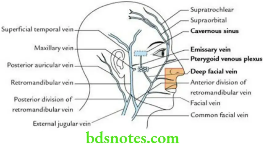
Answer.

External Jugular Vein Formation The external jugular vein is formed on the sternocleidomastoid muscle below and behind the angle of the mandible by the union of the posterior auricular vein and posterior division of the retromandibular vein. It has two valves – one before its termination and another an inch above the middle of the clavicle.

“Best Ways To Understand External Jugular Vein Anatomy”

Head And Neck Scalp temple and face Venous drainage of the face

“Importance Of The External Jugular Vein In Circulation”

External Jugular Vein Course It descends obliquely downwards and backwards across the sternocleidomastoid. It pierces the superficial lamina of the investing layer of deep cervical fascia above the clavicle in the region of the subclavian triangle. Here it crosses the third part of the subclavian artery and after piercing a deep lamina of the investing layer of the deep cervical fascia ends in the subclavian vein deep into the clavicle.

External Jugular Vein Tributaries These are

  • Anterior jugular vein
  • Transverse cervical vein
  • Suprascapular vein

“Risk Factors For External Jugular Vein Complications”

External Jugular Vein Applied anatomy

  • Air embolism: If the external jugular vein is cut an inch above the clavicle, its lumen is held open because its margins are adhered to the deep fascia. As a result, the air is sucked into the lumen of the external jugular vein during inspiration, leading to air embolism that may subsequently cause death.
  • The external jugular vein is used by clinicians to measure the external jugular venous pressure and/or pressure in the right atrium.

Filed Under: Head And Neck

Reader Interactions

Leave a Reply Cancel reply

Your email address will not be published. Required fields are marked *

Primary Sidebar

Recent Posts

  • Branchial Cleft Cyst: Background, Pathophysiology, Etiology
  • Maxillary Nerve: Origin, Course, And Branches
  • The Father Of Anatomy And A Great Anatomist Herophilus
  • Bone Structure – Anatomy
  • The External Carotid Artery: Anatomy, Branches, And Functions
  • Occipitofrontalis Muscle
  • Superficial Temporal Artery
  • Platysma Muscle
  • Cartilage
  • Cauda Equina And Conus Medullaris Syndromes
  • Subcutaneous Injections And Device Management
  • Types Of Circulation: Pulmonary, Systemic, And Portal
  • Structure Of Skeletal Muscle
  • Elastic Cartilage
  • Cellular Organelles And Structure
  • The Golgi Apparatus – The Cell
  • The Cytoplasmic Inclusions Of Certain Plant Cells
  • Dental Abscess
  • Laser Surgery
  • Our Facial Muscles And Their Functions

Copyright © 2026 · Magazine Pro on Genesis Framework · WordPress · Log in