Pterygopalatine Ganglion: Anatomy, Location And Function
Describe the pterygopalatine ganglion (sphenopalatine ganglion) in brief under the following headings:
- Pterygopalatine Ganglion Location,
- Pterygopalatine Ganglion Roots,
- Pterygopalatine Ganglion Branches,
- Pterygopalatine Ganglion Distribution and
- Pterygopalatine Ganglion Applied anatomy.
Answer.
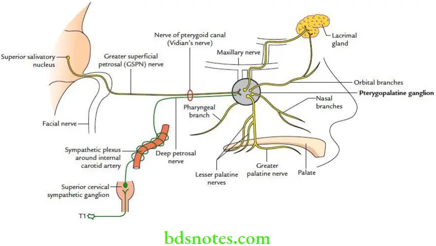
Pterygopalatine Ganglion Location The pterygopalatine ganglion is the largest peripheral parasympathetic ganglion. It is located in the pterygopalatine fossa.
“Importance Of The Pterygopalatine Ganglion In Facial Innervation”
Pterygopalatine Ganglion Roots It has following three roots:
Parasympathetic root: From greater petrosal nerve.
- Sympathetic root: From sympathetic plexus around internal carotid artery through deep petrosal nerve.
- Sensory root: From maxillary nerve.
“Risk Factors For Damage To The Pterygopalatine Ganglion”
Pterygopalatine Ganglion Branches
- Orbital branches
- Palatine branches
- Nasal branches
Pterygopalatine Ganglion Distribution
- Parasympathetic (secretomotor) fibres: Supply the lacrimal, nasal and palatine glands.
- Sympathetic (vasomotor) fibres: Supply the mucous membrane of the nose, paranasal air sinuses and nasopharynx.
- Sensory fibres: Provide sensory innervation to the periosteum of orbit and mucous membrane of nose, palate and pharynx.
“Early Signs Of Issues With The Pterygopalatine Ganglion”
Pterygopalatine Ganglion Applied anatomy
- Allergic conditions such as hay fever or cold cause irritation of the pterygopalatine ganglion, which leads to running of the nose and eyes. For this reason, the pterygopalatine ganglion is also termed the ganglion of hay fever.
- The alcohol injection is occasionally used to relieve/treat intractable cases of allergic rhinitis.
Pterygopalatine Fossa
The pterygopalatine fossa is a pyramidal-shaped space that lies in the depth of the pterygomaxillary fissure.
“The Role Of Imaging In Diagnosing Pterygopalatine Ganglion Disorders”
Question 1. Define pterygopalatine fossa and enumerate its boundaries and contents.
Answer.
Pterygopalatine Fossa Boundaries
Anterior: Posterolateral surface of maxilla.
Posterior: Pterygoid process and greater wing of the sphenoid.
“Understanding The Role Of The Pterygopalatine Ganglion In Facial Innervation”
Medial: Perpendicular plate of palatine.
Lateral: Fossa presents a pterygomaxillary fissure.
Floor: Angle between the anterior and posterior walls of the fossa.
Roof: Body of sphenoid.
“Comprehensive Overview Of The Pterygopalatine Ganglion And Its Significance”
Pterygopalatine Fossa Contents
- Maxillary nerve
- Pterygopalatine ganglion
- Maxillary artery (3rd part)
Leave a Reply