• Skip to main content
  • Skip to secondary menu
  • Skip to primary sidebar
  • About Us
  • Terms of Use
  • Privacy Policy
  • Disclaimer
  • Contact Us
  • Sitemap

BDS Notes

BDS notes, Question and Answers

  • Public Health Dentistry
  • Periodontics
  • Pharmacology
  • Pathology
  • Orthodontics
    • Anchorage In Orthodontics
    • Mandibular Growth, Functional Matrix
    • Retention and Relapse
  • General Surgery
    • Cysts: Types, Causes, Symptoms
    • Maxillofacial Fractures, Disorders, and Treatments
    • Lymphatic Disorders
    • Neurological and Facial Disorders
  • Temporal And Infratemporal Regions
    • Spinal and Neuroanatomy
  • Dental Materials
    • Dental Amalgam
Home » Parotid Gland Comparative Microscopic Anatomy

Parotid Gland Comparative Microscopic Anatomy

February 9, 2026 by Kristensmith Taylor Leave a Comment

Parotid Gland Comparative Microscopic Anatomy

Describe the histological features of the parotid gland in brief.
Answer.

It is a compound tubuloalveolar gland and presents the following histological features.

Head And Neck Parotid and submandibular regions Histological features of partoid salivary gland

Connective tissue:

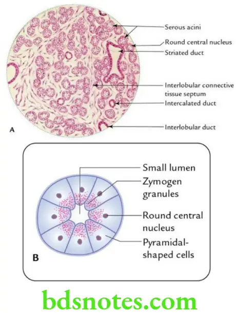
The connective tissue (fibrous) septa divide the gland into lobes and lobules.

Acini:

  • Presence of a large number of serous acini.
  • Acini are round and show biphasic stain with H&E stain, basophilic in basal part and eosinophilic in apical parts. They have very small lumen.
  • Acini are lined by pyramidal cells with round nuclei placed near the centre.

Ducts:

  • Intercalated ducts are lined by simple squamous epithelium.
  • Striated ducts are lined by simple columnar epithelium with basal striations. They are stained dark with eosin.
  • Interlobular ducts are present in connective tissue septa between the lobules and are lined by stratified cuboidal/low columnar epithelium.

Filed Under: Head And Neck

Reader Interactions

Leave a Reply Cancel reply

Your email address will not be published. Required fields are marked *

Primary Sidebar

Recent Posts

  • Branchial Cleft Cyst: Background, Pathophysiology, Etiology
  • Maxillary Nerve: Origin, Course, And Branches
  • The Father Of Anatomy And A Great Anatomist Herophilus
  • Bone Structure – Anatomy
  • The External Carotid Artery: Anatomy, Branches, And Functions
  • Occipitofrontalis Muscle
  • Superficial Temporal Artery
  • Platysma Muscle
  • Cartilage
  • Cauda Equina And Conus Medullaris Syndromes
  • Subcutaneous Injections And Device Management
  • Types Of Circulation: Pulmonary, Systemic, And Portal
  • Structure Of Skeletal Muscle
  • Elastic Cartilage
  • Cellular Organelles And Structure
  • The Golgi Apparatus – The Cell
  • The Cytoplasmic Inclusions Of Certain Plant Cells
  • Dental Abscess
  • Laser Surgery
  • Our Facial Muscles And Their Functions

Copyright © 2026 · Magazine Pro on Genesis Framework · WordPress · Log in