• Skip to main content
  • Skip to secondary menu
  • Skip to primary sidebar
  • About Us
  • Terms of Use
  • Privacy Policy
  • Disclaimer
  • Contact Us
  • Sitemap

BDS Notes

BDS notes, Question and Answers

  • Public Health Dentistry
  • Periodontics
  • Pharmacology
  • Pathology
  • Orthodontics
    • Anchorage In Orthodontics
    • Mandibular Growth, Functional Matrix
    • Retention and Relapse
  • General Surgery
    • Cysts: Types, Causes, Symptoms
    • Maxillofacial Fractures, Disorders, and Treatments
    • Lymphatic Disorders
    • Neurological and Facial Disorders
  • Temporal And Infratemporal Regions
    • Spinal and Neuroanatomy
  • Dental Materials
    • Dental Amalgam
Home » Parotid Gland: Anatomy, Function, Location & Definition

Parotid Gland: Anatomy, Function, Location & Definition

February 9, 2026 by Kristensmith Taylor Leave a Comment

Parotid Gland: Anatomy, Function, Location & Definition

Write a short note on the parotid capsule.
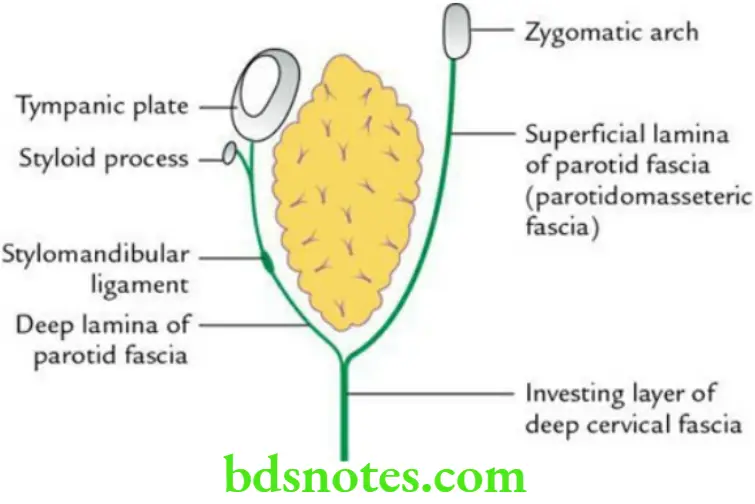
Answer.

It is a facial capsule that encloses the parotid gland. It is derived from the investing layer of the deep cervical fascia. At the lower end of the gland, the fascia splits into superficial and deep laminae to enclose the gland.

  • The superficial lamina is extremely dense and tough and is attached to the lower border of the zygomatic arch. It blends with the perimysium of the masseter to form the parotid masseteric fascia.
  • The deep lamina is relatively thin and is attached to the styloid process, tympanic plate and mandible. It also forms the stylomandibular ligament.

Head And Neck Parotid and submandibular regions Parotid capsule

Filed Under: Head And Neck

Reader Interactions

Leave a Reply Cancel reply

Your email address will not be published. Required fields are marked *

Primary Sidebar

Recent Posts

  • Branchial Cleft Cyst: Background, Pathophysiology, Etiology
  • Maxillary Nerve: Origin, Course, And Branches
  • The Father Of Anatomy And A Great Anatomist Herophilus
  • Bone Structure – Anatomy
  • The External Carotid Artery: Anatomy, Branches, And Functions
  • Occipitofrontalis Muscle
  • Superficial Temporal Artery
  • Platysma Muscle
  • Cartilage
  • Cauda Equina And Conus Medullaris Syndromes
  • Subcutaneous Injections And Device Management
  • Types Of Circulation: Pulmonary, Systemic, And Portal
  • Structure Of Skeletal Muscle
  • Elastic Cartilage
  • Cellular Organelles And Structure
  • The Golgi Apparatus – The Cell
  • The Cytoplasmic Inclusions Of Certain Plant Cells
  • Dental Abscess
  • Laser Surgery
  • Our Facial Muscles And Their Functions

Copyright © 2026 · Magazine Pro on Genesis Framework · WordPress · Log in