Mumps: Causes, Symptoms & Treatments
Describe the parotid gland under the following headings:
- External features,
- Relations,
- Nerve supply and
- Applied anatomy.
Answer.
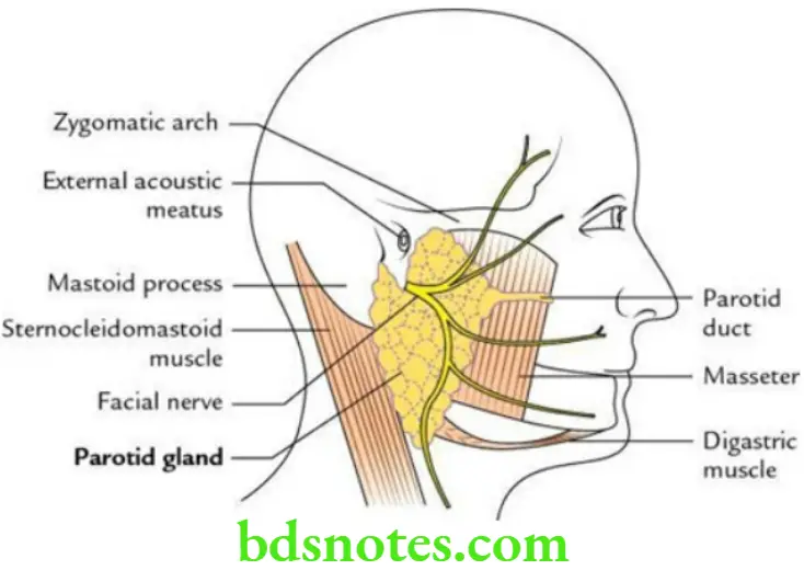
Parotid Gland External features The parotid gland is the largest salivary gland (weight: 25 g) located in the parotid region. It resembles a three-sided inverted pyramid and presents the following features.
Apex: Directed downwards
Base: Directed upwards
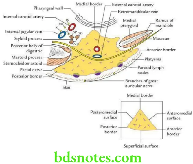
Parotid Gland Three surfaces:
- Superficial (largest)
- Anteromedial
- Postmedial
Parotid Gland Three borders:
- Anterior
- Posterior
- Medial
Read And Learn More: Selective Anatomy Notes And Question And Answers
Relations
Apex: It is related to the posterior belly of the digastric.
Base: It is related to:
- External acoustic meatus
- Posterior aspect of the temporomandibular joint
Superficial surface: It is related to:
- Skin
- Superficial fascia containing platysma, branches of great auricular nerve and superficial parotid lymph nodes
- Parotidomasseteric fascia
- Deep parotid lymph nodes embedded in the gland
Anteromedial surface: It is related to:
- Masseter
- Posterior border of ramus of mandible
- Medial pterygoid
Posteromedial surface: It is related to:
- Mastoid process and muscles covering it
- Styloid process and muscles covering it
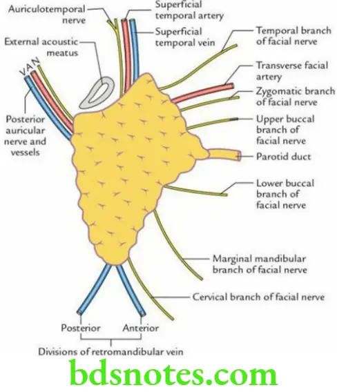
Anterior border: The following structures (from above to downwards) emerge underneath this border:
Posterior border: The following structures emerge from underneath this border:
- Posterior auricular nerve
- Posterior auricular vessels
Medial border: It is related to the lateral wall of the pharynx.
Nerve supply
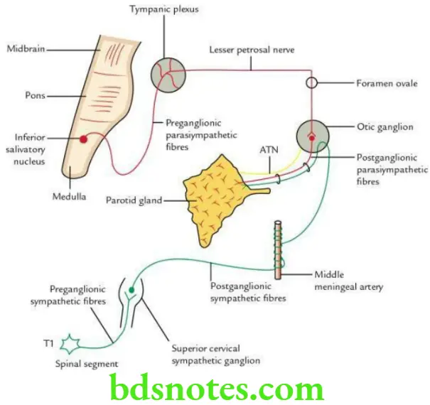
Secretomotor (parasympathetic): Preganglionic fibres arising from the inferior salivatory nucleus and travel to the otic ganglion as follows:
Inferior salivatory nucleus → Glossopharyngeal nerve → Tympanic branch of glossopharyngeal nerve (Jacobson’s nerve) → Tympanic plexus → Lesser petrosal nerve → Relay in otic ganglion
Postganglionic fibres arise from otic ganglion and travel through an auriculotemporal nerve to supply the parotid gland.
Vasomotor (sympathetic):
- Preganglionic fibres arise from the T1 segment of the spinal cord and relay in the superior cervical sympathetic ganglion.
- Postganglionic fibres arise from the superior cervical sympathetic ganglion and run along the arteries (e.g. external carotid artery) to supply the gland.
Vasomotor Sensory:
- Auriculotemporal nerve
- Great auricular nerve
Vasomotor Applied anatomy
Mumps (viral parotitis): It is the inflammation of the parotid gland by the mumps virus. Mumps characteristically do not suppurate. In adults, the mumps may cause complications like orchitis in males, oophoritis in females and pancreatitis in both sexes.
Parotid swellings: These are very painful due to the unyielding nature of parotid fascia, which encloses the gland.
Parotid abscess: It is drained by a transverse incision in the parotid masseteric fascia to avoid injuries to the facial nerve (Hilton’s method).
Frey syndrome: It occurs due to the damage of branches of the auriculotemporal and great auricular nerves by penetrating wounds in the parotid region. During regeneration, secretomotor fibres of the auriculotemporal nerve join the fibres of the great auricular nerve.
As a result, the stimulation of the parotid gland causes stimulation of the great auricular nerve, which leads to sweating and redness (hyperaemia) in the area of distribution of the great auricular nerve (e.g. parotid region).
Leave a Reply