• Skip to main content
  • Skip to secondary menu
  • Skip to primary sidebar
  • About Us
  • Terms of Use
  • Privacy Policy
  • Disclaimer
  • Contact Us
  • Sitemap

BDS Notes

BDS notes, Question and Answers

  • Public Health Dentistry
  • Periodontics
  • Pharmacology
  • Pathology
  • Orthodontics
    • Anchorage In Orthodontics
    • Mandibular Growth, Functional Matrix
    • Retention and Relapse
  • General Surgery
    • Cysts: Types, Causes, Symptoms
    • Maxillofacial Fractures, Disorders, and Treatments
    • Lymphatic Disorders
    • Neurological and Facial Disorders
  • Temporal And Infratemporal Regions
    • Spinal and Neuroanatomy
  • Dental Materials
    • Dental Amalgam
Home » Extraocular Muscles: Anatomy And Movements

Extraocular Muscles: Anatomy And Movements

February 5, 2025 by Kristensmith Taylor Leave a Comment

Extraocular Muscles: Anatomy And Movements

Write about the origin, insertion, nerve supply and actions of the extrinsic muscles of the eyeball.
Answer.

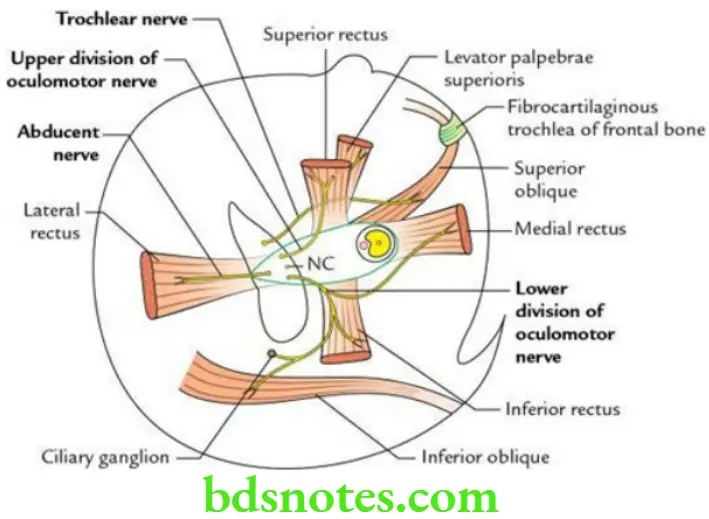
Extrinsic Muscles of Eyeball Origin

Four recti: From common tendinous ring.

Superior oblique: From the body of the sphenoid superomedial to the optic canal.

Inferior oblique: From a rough impression in the anteromedial part/angle of the floor of the orbit.

“Understanding the anatomy and movements of extraocular muscles through FAQs: Q&A explained”


Head And Neck Ear and orbit Origin and nerve supply of extraocular muscles

“Importance of studying extraocular muscles for medical students: Questions explained”

Extrinsic Muscles of Eyeball  Insertion

Four recti: Into sclera, a little posterior to the limbus (i.e. corneoscleral junction). Distance from the cornea is as follows:

SR = 7.7 mm; IR = 6.5 mm; MR = 5.5 mm; LR = 6.9 mm

Superior oblique: Into sclera, behind the equator in the posterosuperior quadrant of the eyeball between SR and LR.

Inferior oblique: Into the sclera, behind the equator in the posteroinferior quadrant of the eyeball.

“Common challenges in understanding extraocular muscle anatomy effectively: FAQs provided”

Extrinsic Muscles of the Eyeball  Nerve supply

All the extrinsic muscles of the eyeball are supplied by CN 3, except the superior oblique, which is supplied by CN 4 and the lateral rectus which is supplied by CN 6.

Mnemonic: LR6SO4 (LR, lateral rectus; SO, superior oblique)

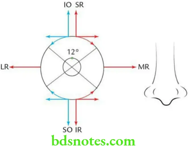
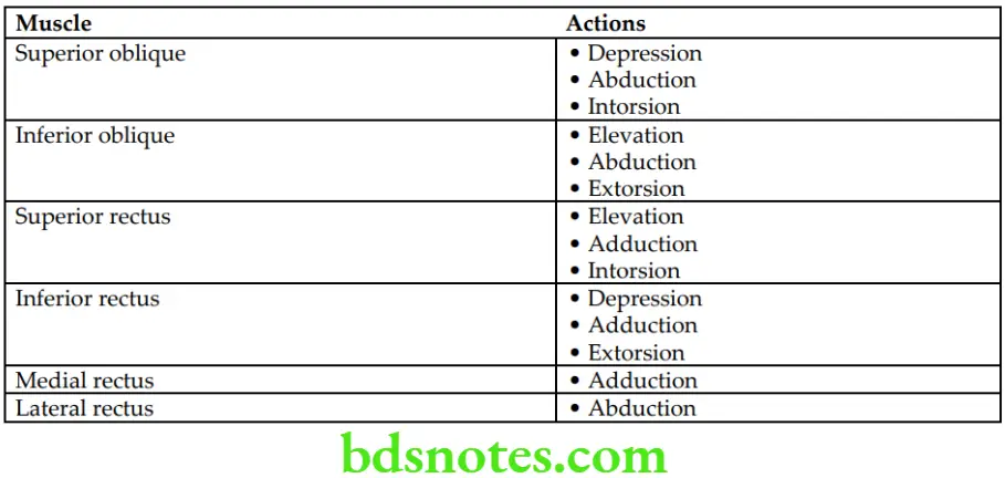
Extrinsic Muscles of Eyeball  Actions

“Role of the lateral rectus muscle in eye abduction: Questions answered”

Head And Neck Ear and orbit Schematic diagram source

“Factors influencing success with extraocular muscle knowledge: Q&A”

Actions of Extrinsic Muscles of the Eyeball

Head And Neck Ear and orbit Actions of extrinsic muscles of the eyeball

“Steps to explain the anatomy of extraocular muscles: Origins vs insertions vs innervation: Q&A guide”

Filed Under: Head And Neck

Reader Interactions

Leave a Reply Cancel reply

Your email address will not be published. Required fields are marked *

Primary Sidebar

Recent Posts

  • Branchial Cleft Cyst: Background, Pathophysiology, Etiology
  • Maxillary Nerve: Origin, Course, And Branches
  • The Father Of Anatomy And A Great Anatomist Herophilus
  • Bone Structure – Anatomy
  • The External Carotid Artery: Anatomy, Branches, And Functions
  • Occipitofrontalis Muscle
  • Superficial Temporal Artery
  • Platysma Muscle
  • Cartilage
  • Cauda Equina And Conus Medullaris Syndromes
  • Subcutaneous Injections And Device Management
  • Types Of Circulation: Pulmonary, Systemic, And Portal
  • Structure Of Skeletal Muscle
  • Elastic Cartilage
  • Cellular Organelles And Structure
  • The Golgi Apparatus – The Cell
  • The Cytoplasmic Inclusions Of Certain Plant Cells
  • Dental Abscess
  • Laser Surgery
  • Our Facial Muscles And Their Functions

Copyright © 2026 · Magazine Pro on Genesis Framework · WordPress · Log in