Pituitary Gland – Definition, Anatomy, Hormones, & Disorders
Describe the pituitary gland under the following headings:
- Pituitary Gland Definition and location,
- Pituitary Gland Gross features,
- Pituitary Gland Relations,
- Pituitary Gland Microscopic structure,
- Pituitary Gland Blood supply,
- Pituitary Gland Nerve supply,
- Pituitary Gland Development and
- Pituitary Gland Applied anatomy.
Answer.
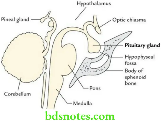
Definition and location Pituitary Gland: The pituitary gland/hypophysis cerebri is a small neuroendocrine gland located in the hypophyseal fossa (sella turcica) of the body of the sphenoid.
“Understanding the anatomy and function of the pituitary gland through FAQs: Q&A explained”
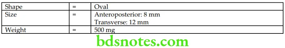
Pituitary Gland Shape and Measurements
Pituitary Gland Gross Features
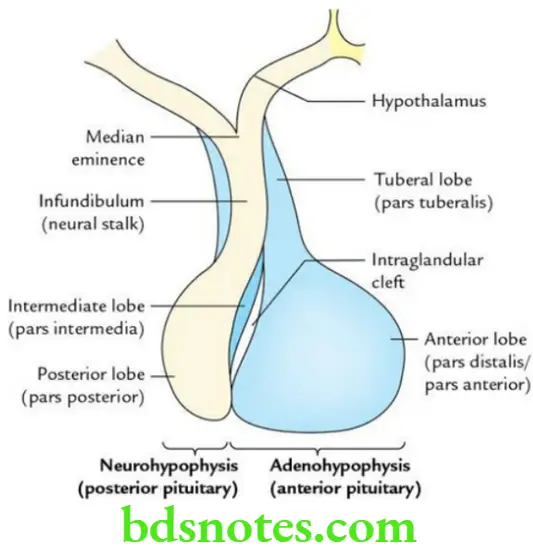
The pituitary gland consists of two parts, which are embryologically, morphologically and functionally different from each other:
- Adenohypophysis (anterior pituitary)
- Neurohypophysis (posterior pituitary)
“Importance of studying the pituitary gland for medical students: Questions explained”
“Common challenges in understanding pituitary gland anatomy effectively: FAQs provided”
Adenohypophysis (Anterior Pituitary)
It is highly cellular and presents an intraglandular cleft. It is further subdivided into three parts/lobes:
- Anterior lobe (pars distalis): It is the major part of the adenohypophysis.
- Intermediate lobe (pars intermedia): It is a thin strip of glandular tissue between the intraglandular cleft in front and neurohypophysis behind.
- Tuberal lobe (pars tuberalis): It is an upward extension of the anterior lobe that surrounds the part of the infundibulum.
Neurohypophysis (Posterior Pituitary):
It is continuous above the infundibulum, which extends downward and forward from the floor of the 3rd ventricle and enters the hypophyseal fossa after piercing the diaphragm sellae. It is subdivided into three parts:
- Posterior lobe (pars posterior): It is smaller than the anterior lobe and lies in the posterior concavity of the larger anterior lobe.
- Infundibulum (neural stalk): It contains the neural connections of the neurohypophysis.
- Median eminence of the tuber cinereum: It is continuous with the infundibular stem.
“Factors influencing success with pituitary gland knowledge: Q&A”
Pituitary Gland Relations
Anterior:
- Anterior intercavernous sinus
Posterior:
- Posterior intercavernous sinus
- Dorsum sellae
- Basilar artery
- Pons
Superior:
- Diaphragma sellae
- Optic chiasma
- Tuber cinereum
- Infundibular recess of the 3rd ventricle
“Steps to apply pituitary gland knowledge in clinical practice: Diagnosis vs treatment: Q&A guide”
Inferior:
- Hypophyseal fossa
- Body of the sphenoid
- Sphenoidal air sinuses
Lateral (on each side):
- Cavernous sinus with its contents
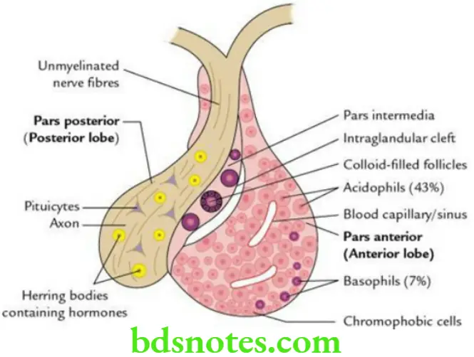
Pituitary Gland Microscopic Structure
Anterior lobe (pars anterior/distal): It forms three-fourths of the gland. It consists of two types of glandular cells arranged in irregular cords or clumps:
- Chromophilic cells (50%) having affinity to colours, i.e. staining
- Acidophils/alpha cells (about 43% of cells): Secrete GH, ACTH and prolactin
- Basophils/beta cells (about 7% of cells): Secrete TSH, LH, FSH and MSH
- Chromophobic cells (50%), which do not take the colour of stain: Resting cells, no secretion
“Role of imaging techniques in diagnosing pituitary gland disorders: Questions answered”
“Early warning signs of complications from ignoring pituitary gland care: Common questions”
Intermediate lobe (pars intermedia): It is made up of numerous basophilic and chromophobic cells. The cells are arranged in the form of small follicles containing colloid material.
Posterior lobe (pars posterior/nervosa): It consists of:
- A large number of nonmyelinated nerve fibres.
- Modified neuroglial cells called pituicytes.
- Presence of Herring bodies, the small, spherical masses that stain deeply with chrome–alum haematoxylin.
“Differential applications of conservative vs surgical treatments for pituitary gland issues: Q&A”
Pituitary Gland Arterial Supply
Venous drainage: Small short veins emerge from the surface of the gland and drain into neighbouring dural venous sinuses.
Pituitary Gland Nerve supply: By hypothalamo-hypophyseal tract, which arises from preoptic and paraventricular nuclei of the hypothalamus.
Pituitary Gland Development: The pituitary gland develops from two distinct sources:
- Adenohypophysis develops from Rathke’s pouch – an ectodermal diverticulum (outpocketing) from the roof of the stomodeum that grows cranially in front of the buccopharyngeal membrane.
- Neurohypophysis develops from the infundibulum – a down growth from the floor of 3rd ventricle.
“Asymptomatic vs symptomatic effects of outdated pituitary gland practices: Answered”
Pituitary Gland Applied Anatomy
The pituitary gland produces a number of hormones that control the secretions of other endocrine glands of the body.
- Pituitary adenoma: It is not uncommon. It compresses the central part of optic chiasma leading to bitemporal hemianopia (tunnel vision).
- Gigantism and acromegaly: Gigantism occurs due to excessive secretion of growth hormone (GH) before adolescence; hence, the person becomes very tall (8–9 feet) due to excessive length of the bone. The acromegaly occurs due to excessive secretion of GH in adults leading to coarse facial features with protrusion of jaw (prognathism).
- Pituitary dwarfism: Occurs due to hyposecretion of GH causing shortening of limbs.
Leave a Reply