• Skip to main content
  • Skip to secondary menu
  • Skip to primary sidebar
  • About Us
  • Terms of Use
  • Privacy Policy
  • Disclaimer
  • Contact Us
  • Sitemap

BDS Notes

BDS notes, Question and Answers

  • Public Health Dentistry
  • Periodontics
  • Pharmacology
  • Pathology
  • Orthodontics
    • Anchorage In Orthodontics
    • Mandibular Growth, Functional Matrix
    • Retention and Relapse
  • General Surgery
    • Cysts: Types, Causes, Symptoms
    • Maxillofacial Fractures, Disorders, and Treatments
    • Lymphatic Disorders
    • Neurological and Facial Disorders
  • Temporal And Infratemporal Regions
    • Spinal and Neuroanatomy
  • Dental Materials
    • Dental Amalgam
Home » The Scalene Muscles – Attachments – Action – Innervation

The Scalene Muscles – Attachments – Action – Innervation

February 7, 2025 by Kristensmith Taylor Leave a Comment

The Scalene Muscles – Attachments – Action – Innervation

Describe the Scalenus Medius Muscle in brief.
Answer.

Scalene Muscles is the longest and largest scalene muscle.

“Early Signs Of Scalene Muscle Dysfunction”

Head And Neck Deep structures of the neck and prevertebral region Attachments of scalenus medius muscle

“Importance Of Scalene Muscles In Neck Movement”

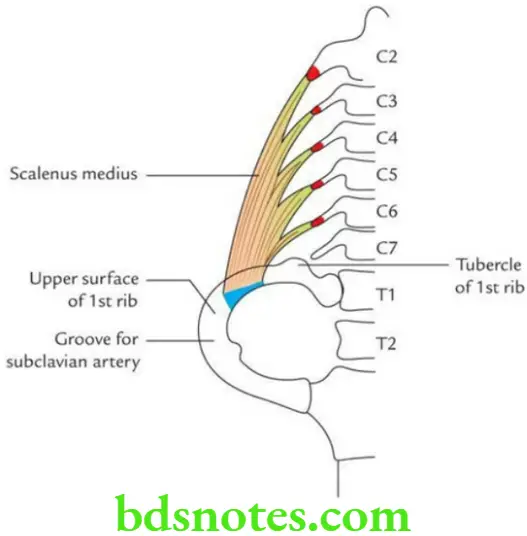
Scalenus Medius Muscle  Origin

From the posterior tubercles of transverse processes of C2 to C6 vertebrae.

Scalenus Medius Muscle  Insertion

On the superior surface of 1st rib behind the groove for subclavian artery and in front of tubercle of 1st rib.

Scalenus Medius Muscle  Nerve supply

Ventral rami of C3 to C8 spinal nerves.

Scalenus Medius Muscle  Actions

Same as scalenus anterior.

Filed Under: Head And Neck

Reader Interactions

Leave a Reply Cancel reply

Your email address will not be published. Required fields are marked *

Primary Sidebar

Recent Posts

  • Branchial Cleft Cyst: Background, Pathophysiology, Etiology
  • Maxillary Nerve: Origin, Course, And Branches
  • The Father Of Anatomy And A Great Anatomist Herophilus
  • Bone Structure – Anatomy
  • The External Carotid Artery: Anatomy, Branches, And Functions
  • Occipitofrontalis Muscle
  • Superficial Temporal Artery
  • Platysma Muscle
  • Cartilage
  • Cauda Equina And Conus Medullaris Syndromes
  • Subcutaneous Injections And Device Management
  • Types Of Circulation: Pulmonary, Systemic, And Portal
  • Hierarchical Organization Of Skeletal Muscle Tissue
  • Elastic Cartilage Histology Short Note For Medical Exams
  • Cellular Organelles And Structure
  • The Golgi Apparatus – The Cell
  • The Cytoplasmic Inclusions Of Certain Plant Cells
  • Dental Abscess
  • Laser Surgery
  • Our Facial Muscles And Their Functions

Copyright © 2026 · Magazine Pro on Genesis Framework · WordPress · Log in