Elbow Joint Anatomy: Classification, Ligaments, Movements
Describe the elbow joint under the following headings: (a) classification, (b) articular surfaces, (c) ligaments, (d) relations, (e) nerve supply, (f) movements, and (g) applied anatomy.
Answer.
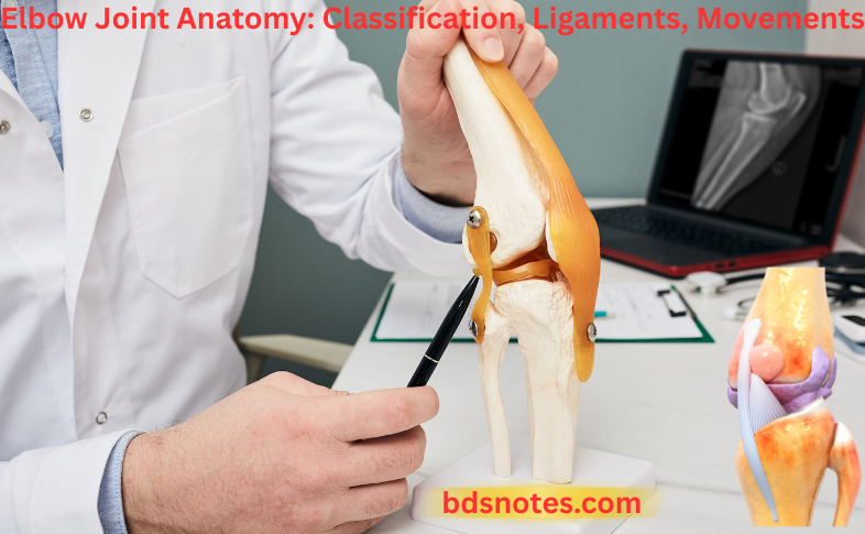
Elbow Joint Classification
Synovial joint of hinge variety.
Articular surfaces
It is a compound joint consisting of two articulations.
Humeroradial: Between capitulum of humerus and head of radius.
Humeroulnar: Between the trochlea of the humerus and the trochlear notch of the ulna.
Elbow Joint Anatomy: Classification, Ligaments, and Movements
Read And Learn More: Selective Anatomy Notes And Questions, And Answers
“Role of surgery in repairing severe ligament tears”

“Understanding the classification of the elbow joint”
The elbow joint communicates with the superior radioulnar joint.
Elbow Joint Ligaments
Elbow Joint Capsular Ligament
- Attachments
- In front
- Superiorly, it is attached to the humerus above the coronoid and radial fossae.
- Inferiorly, it is attached to the coronoid process of the ulna and the annular ligament of the superior radioulnar joint.
- Behind
- Superiorly, it is attached to the margins of the olecranon fossa.
- Internally, it is attached to the upper margins of the olecranon process and the annular ligament of a superior radioulnar joint.
- In front
- On either side, the capsule becomes continuous with the medial and lateral collateral ligaments of the elbow joint.
Elbow Joint Anatomy and Function
Elbow Joint Medial (ulnar) collateral ligament:
Elbow Joint Medial (ulnar) collateral ligament is triangular and consists of the following three parts:
- Anterior part, which extends from the front of the medial epicondyle of humerus to the medial margin of the coronoid process of the ulna
- Posterior part, which extends from the back of the medial epicondyle of humerus to the medial margin of the olecranon process of the ulna
- Inferior part, which extends between the lower ends of the anterior and posterior parts and stretches between the olecranon and coronoid processes of the ulna
“Treatment options for common elbow joint injuries”
“Importance of studying elbow joint anatomy for healthcare professionals”
Lateral (radial) collateral ligament:
Elbow Joint Lateral (radial) collateral ligament extends from the lateral epicondyle of the humerus above to the annular ligament below.
Elbow Joint Relations
Elbow Joint Anterior
- Brachialis
- Tendon of biceps brachii
- Median nerve
- Brachial artery
Elbow Joint Posterior
- Tendon of triceps brachii
- Anconeus
Elbow Joint Medial
- Flexor carpi ulnaris
- Ulnar nerve
- Common flexor origin
“Global prevalence of elbow joint injuries in athletes”
Elbow Joint Lateral
- Supinator
- Common extensor origin
Elbow Joint Nerve Supply
- Radial nerve
- Musculocutaneous nerve
- Median nerve
- Ulnar nerve
Elbow Joint Movements
The movements and muscles producing them, with their nerve supply.
Movements of the Elbow Joint and Muscles Producing Them with Their Nerve Supply

“Case studies on outcomes of elbow joint injury treatments”
Elbow Joint Applied Anatomy
Elbow Joint Dislocation:
The dislocation of the elbow joint usually occurs posteriorly and is often associated with a fracture of the coronoid process.
Elbow Joint Effusion:
The effusion of the elbow joint causes distension on the posterior aspect of the elbow because the joint capsule is weak posteriorly.
Elbow Joint Ligaments and Movements
Tennis elbow (lateral epicondylitis)
Tennis Elbow presents with pain and tenderness over the lateral epicondyle of the humerus. It occurs due to:
- Sprain of the lateral collateral ligament of the elbow joint
- Tearing of the fibres of the extensor carpi radialis brevis (ECRB)
- Inflammation of the bursa underneath the ECRB
- Tear of the common extensor origin
Leave a Reply