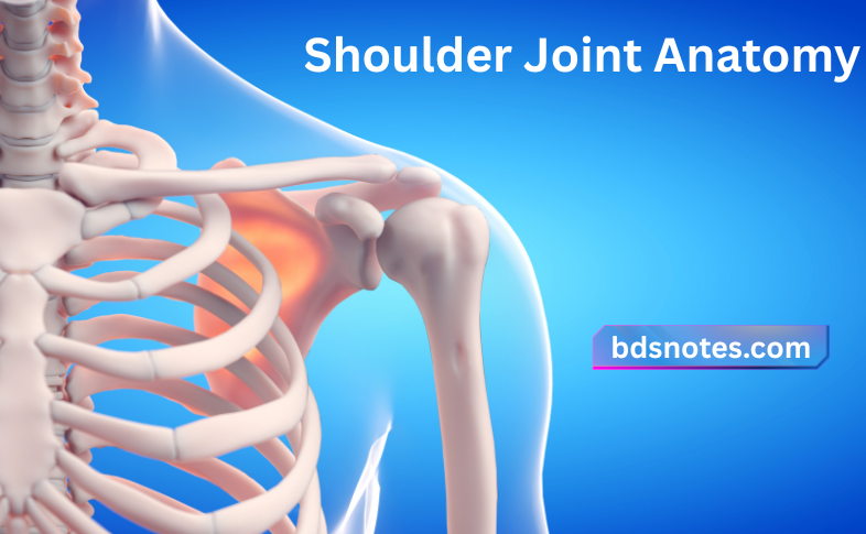
Shoulder Joint Anatomy
Question 1. Describe the Shoulder Joint (Glenohumeral Joint) under the following headings: (a) classification, (b) articular surfaces, (c) ligaments, (d) relations, (e) nerve supply, (f) movements and (g) applied anatomy.
Answer.
Shoulder Joint (Glenohumeral Joint) Classification
Synovial joint of ball and socket type.
Shoulder Joint (Glenohumeral Joint) Articular Surfaces
They are formed by the large hemispherical head of the humerus and the shallow glenoid cavity of the scapula.
Shoulder Joint Anatomy
Read And Learn More: Selective Anatomy Notes And Questions And Answers
“Understanding the structure and function of the shoulder joint”
“Importance of studying shoulder joint anatomy for healthcare professionals”
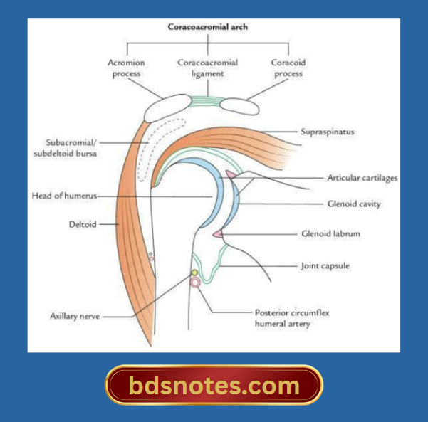
The glenoid articular surface is deepened by the glenoid labrum.
Glenoid labrum
- It is a rim of fibrocartilage attached to the peripheral margin of the glenoid cavity.
- It is triangular in cross-section and deepens the shallow glenoid cavity.
Shoulder Joint Ligaments
Capsular ligament
- Attachments
- Medially: To the peripheral margin of the glenoid cavity outside the glenoid labrum. The supraglenoid tubercle is intracapsular.
- Laterally: To the anatomical neck of the humerus except on the medial side where it descends about 2–3 cm on the shaft, up to the surgical neck of humerus.
- Muscles strengthening the capsule: In general, the capsule is loose and lax, but it is strengthened by the musculotendinous (rotator) cuff formed by the following muscles:
- Openings in the capsule: The capsule presents two openings/deficiencies:
- One in front, for communicating with subscapular bursa
- One at the intertubercular sulcus to provide the passage for the tendon of long head of biceps brachii
Anatomy of the Shoulder Joint
“Role of the glenoid cavity and humeral head in shoulder movement”
Transverse humeral ligament: This ligament bridges across the bicipital groove.
“Treatment options for common shoulder joint injuries”
Glenohumeral ligaments: These are thickenings in the anterior part of the capsule and are seen when the capsule is exposed from behind. They are three in number and named superior, middle, and inferior glenohumeral ligaments according to their location.
Coracohumeral ligament: It is a wide, strong fibrous band on the superior surface of the joint, extending from the base of the coracoid process to the anterior aspect of the greater tubercle of the humerus.
Coracoacromial ligament: It extends between the lateral side of the coracoid process and the medial border of the acromion.
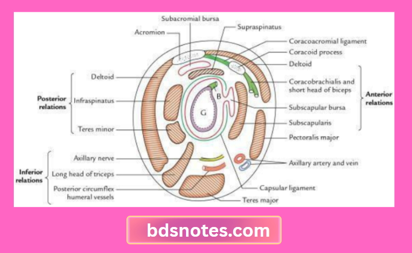
Shoulder Joint (Glenohumeral Joint) Relations
Shoulder Joint Superiorly
- Coracoacromial arch
- Subacromial bursa
- Supraspinatus
- Tendon of the long head of the biceps brachii (intracapsular)
- Deltoid
“Role of surgery in repairing severe rotator cuff tears”
“Follow-up care after addressing shoulder joint dysfunction”
Shoulder Joint Anteriorly
- Subscapularis
- Coracobrachialis
- Short head of biceps
- Deltoid
Shoulder Joint Posteriorly
- Infraspinatus
- Teres minor
- Deltoid
Shoulder Joint Inferiorly
- Long head of triceps
- Axillary nerve
- Posterior circumflex humeral vessels
Shoulder Joint Nerve Supply
The nerves supplying the joint are
- Suprascapular nerve
- Axillary nerve
- Musculocutaneous nerve
Shoulder Joint Movements
The movements and muscles producing them, with their nerve supply.
Movements of the Shoulder Joint and Muscles Producing Them with Their Nerve Supply
“Techniques for managing high-risk groups with injuries”

“Complications of ignoring shoulder joint injuries”
Shoulder Joint (Glenohumeral Joint) Applied Anatomy
Dislocation of the shoulder joint
- The shoulder joint is the most commonly dislocated in the body due to (i) disproportionate size of articular surfaces – head of humerus and glenoid cavity of the scapula (the head of humerus is much larger to fit properly into the smaller glenoid cavity of the scapula [4:1 ratio]) and (ii) laxity of joint capsule.
- Dislocation most commonly occurs inferiorly because the joint is least supported below.
Shoulder Joint Structure and Function
Frozen shoulder (adhesive capsulitis)
It is a clinical condition characterized by painful and uniform restriction of all movements of the shoulder joint. It occurs due to the shrinkage of the joint capsule, leading to adhesion between rotator cuff and the head of the humerus.
Leave a Reply